Diagnosing Anaplasmosis
Veterinarians explain pros and cons of the three main ways of diagnosing anaplasmosis in cattle.

The deadly bacterial disease, bovine anaplasmosis, has wreaked havoc on cattle herds throughout the Southeast. Sporadic incidents of the disease have occurred in other parts of the country.
Mainly infecting ruminants, anaplasmosis attacks the animal’s red blood cells. The body’s built-in defense generates an immune response that attempts to rapidly remove the infected cells via the spleen.
Research discovered an infected animal could have only half the normal amount of red blood cells in circulation. With significantly fewer red blood cells, the animal will become anemic and usually express depressed and lethargic behavior. This behavior leads to weight loss, abortions and sometimes death.
Although some animals’ immune response is strong enough to survive the initial infection, they will become potential carriers and could show reoccurring symptoms for the rest of their lives. Since an immune response is the main control, it is a distinctive trait that anaplasmosis affects older animals as their response may be slower or less effective.
Diagnostic tools
The common acute symptoms presented by an infected animal make the disease difficult to diagnose with the naked eye, said Gregg Hanzlicek, a faculty member at the Kansas State Veterinary Diagnostic Laboratory (KSVDL).
The KSVDL uses three primary practices to diagnose anaplasmosis: blood microscopic exam, serum enzyme-linked immunosorbent assay (ELISA) and polymerase chain reaction (PCR) test. Whether each exam will detect anaplasmosis depends on when the bacterial infection may have occurred, noted Hanzlicek.
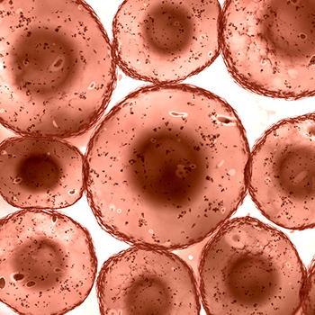
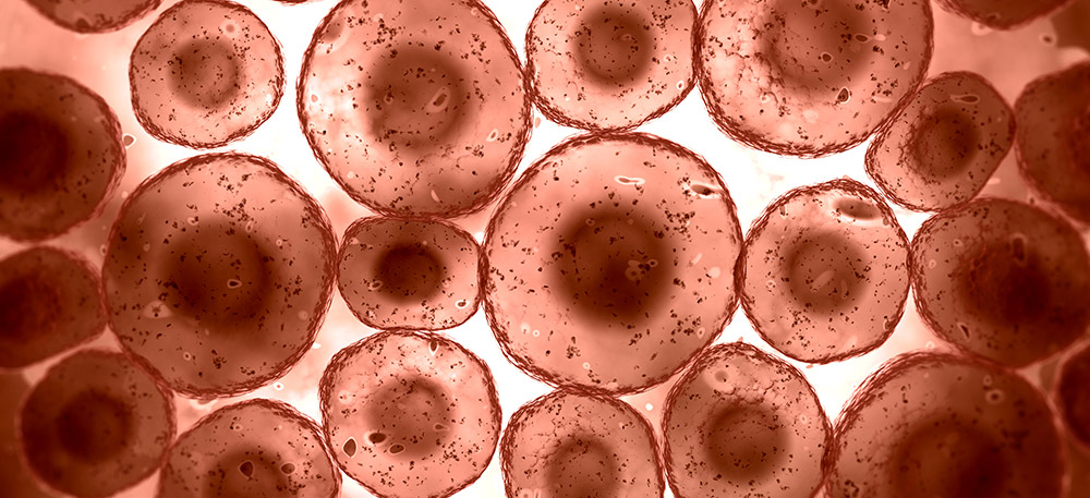
A simple blood sample can be used to create a blood smear to be analyzed under a microscope. Breeders should use this tool to confirm clinical signs of anaplasmosis, which usually occur three weeks after infection.
“If a veterinarian decides to do a microscopic examination, they take a drop of blood, smear it on a microscope slide so it’s one layer thick and stain it,” said Hanzlicek. “An acutely infected animal with anaplasmosis will have irregular-shaped red blood cells.”
A simple blood sample can be used to create a blood smear to be analyzed under a microscope. Breeders should use this tool to confirm clinical signs of anaplasmosis, which usually occur three weeks after infection.
Sampling blood before or after clinical signs appear will not likely result in a positive diagnosis, since the amount of bacteria may be too low to accurately detect. Furthermore, Hanzlicek shared research saying sensitivity is only 20%-26% when trying to detect anaplasmosis in persistently infected animals that have cycles of bacteria growth.
The ELISA test relies on certain antibodies produced by an animal to diagnose anaplasmosis. Due to the cyclical nature of the infection, the ELISA test is less useful when evaluating the disease in its early stages.
“When an animal becomes infected with anaplasmosis, it mounts an immune response and starts building antibodies,” said Hanzlicek. “We take a serum sample to search for those antibodies related to anaplasmosis. This particular test is specifically targeting the bacteria, Anaplasma marginale.”
In most instances the test will be positive for infected animals, vaccinated animals or carriers of anaplasmosis. After a few weeks of the initial infection, the animal will have produced antibodies in an attempt to fight the bacteria, and the ELISA will be positive. Since vaccinated animals were exposed to the bacteria, they will result in a positive outcome, as well. Additionally, previously infected carriers will have the particular antibodies in their blood.
“In a 2007 study that infected 10 cows, it was determined the sensitivity of the ELISA test within 10 days of the initial infection was only 50% sensitivity,” said Hanzlicek. “After enough time was given for those animals to build antibodies up, the test becomes very accurate with a 99.6% sensitivity. There is no test that is 100% accurate, but at the right time the ELISA test is very good for detecting the disease.”
The final tool is a PCR test, which detects the actual bacterial organism from a non-clotted blood sample typically used for necropsied animals. A PCR can detect the multiple strains of anaplasmosis bacteria whether the strain is disease-causing in cattle or not, stated Hanzlicek.
Digging into the results
Along with a test result of positive or negative, the ELISA and PCR tests offer a number value.
The number with an ELISA test is known as an inhibition (I) number that gives a probability of the test’s accuracy. Typically I numbers greater than 30 indicate disease.
“There is a misconception that the I number can also give some prognosis or say whether the animal is in acute infection or not. It doesn’t do any of that,” said Hanzlicek. “It does show the further the number is away from the cutoff, the more credence you can put in that it is actually a true positive result. A negative works the same way. An animal that is negatively far away from that cutoff point indicates we are very sure she is a true negative.”
Animals that result in a number close to the cutoff value are likely to be misclassified as false positives or negatives. Hanzlicek recommended retesting those animals with a PCR or later with another ELISA test.
A PCR test will generate a cycle time value (CT value) that indicates how many organisms are in the animal. Lower CT values specify a higher amount of organisms in the animal.
“When we get numbers down in the twenties or teens, there is a boatload of those organisms in the animal,” said Hanzlicek. “We are going to feel more confident that the disease-causing anaplasmosis organism had something to do with whatever the disease process we are looking at.”
Hanzlicek said the tests are very accurate when used at the correct times, but analyzing the numbers along with a positive or negative outcome will help increase a breeder’s confidence in the results.
If a breeder suspects an animal may have anaplasmosis or is exhibiting similar symptoms, they should contact their veterinarian.
Editor’s note: Kaci Foraker was the 2019 Angus Media summer intern. She is a student at Kansas State University.